Clinical History
A 5-year-old female presented with a complaint of urinary retention.
Ultrasound examination revealed a large, well-defined heterogeneous pelvic lesion measuring 11.7 × 9.8 × 6.6 cm, demonstrating internal vascularity. The lesion was noted to be displacing both ovaries and the urinary bladder superiorly.
Serum Alpha-Fetoprotein (AFP) and Beta Human Chorionic Gonadotropin (β-HCG) levels were within normal limits.
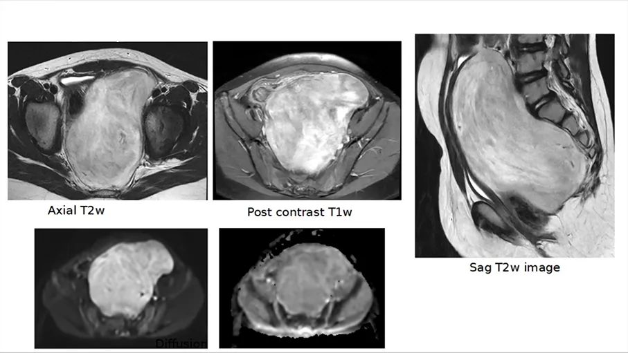
What are the differential diagnosis
1.Rhabdomyosarcoma
2.Ewings sarcoma
3.Germ cell tumour
4.Ganglioneuroma
Diagnosis Answer
Findings:
Location of the mass was presacral. Urinary bladder was displaced anteriorly.
Rectum displaced anteriorly and to the right ( arrow).
Bone was not involved. No extension to the sacral foramina seen.
Biopsy was performed- s/o Ganglioneuroma
Common Differential diagnosis of a presacral mass in this age are:
Congenital- cystic hamartoma, duplication cyst, dermoid / germ cell tumour or anterior meningocele
Osseous- Ewing’s sarcoma, ABC
Mesenchymal- sarcoma
Neurogenic masses- neuroblastoma, ganglioneuroma, ganglioneuroblastoma, nerve sheath tumour
Others- DSRCT, lymphoma.
Ganglion Cell or Neuroblastic Tumors
Neuroblastoma, ganglioneuroblastoma, and ganglioneuroma arise from neural crest cells - from sympathetic chain – and can be located from neck to pelvis & adrenal medulla.
Primordial neural crest cells may remain undifferentiated and immature ( in neuroblastoma) or undergo differentiation into mature ganglion or & constitute ganglioneuromas. Ganglioneuroblastoma contains both differentiated and undifferentiated cells.
Neuroblastoma- 10% of all pediatric cancers and is the most common malignancy in infancy (the 1st year of life).
Common sites are - adrenal gland and retroperitoneum (70%), posterior mediastinum (20% ), neck ( 5%) and pelvis (2%–3%).
Clinical manifestations: Child may present with constitutional symptoms such as fever, weight loss, malaise, and failure to thrive in neuroblastoma. Symptoms secondary to mass effect are known- urinary retention or frequency of evacuation, and constipation.
Radiographic findings - are nonspecific, may have calcification ( stippled in adrenal lesions) in 30%. It may extend into the neural foramina and cause bone remodelling or erosion. It may contain areas of hemorrhage or necrosis and be heterogeneous in attenuation. Smaller tumours may be heterogeneous.
Ganglioneuroma – rare benign tumor, may arise from regressed neuroblastoma / ganglioneuroblastoma or de novo.
More common in older children
Site- posterior mediastinum, retroperitoneum, adrenal gland, and neck, pelvis
Imaging characteristics – No specific imaging features, but tends to be more homogeneous than neuroblastoma and ganglioneuroblastoma. Diagnosis is based on biopsy.
The differential diagnosis
1.Rhabdomyosarcoma and Ewing’s sarcoma - Usually more heterogeneous, may have restricted diffusion. The pelvis is a less common site. Ewing’s sarcoma can be osseous or extra-osseous.
2.Germ cell tumour – May have elevated AFP or b–HCG, or imaging may show fat component, cystic areas, or calcification.
3.Ganglioneuroma – Was more in favor due to relatively homogeneous appearance on T2w images